
Spinal angiograms are usually performed to search for and to localise an arteriovenous malformation (AVM) or fistula in or around the spinal cord. These encompass different forms of abnormal connections between the arteries supplying the spinal cord and its coverings and draining veins. They are broadly classified into dural fistulas, epidural fistulas, perimedullary fistulas and spinal cord AVMs.
A complete spinal angiogram involves selective catheterisation of numerous arteries on each side of the spine as well as in the pelvis and neck. The spinal arteries are small and optimal visualisation may require immobilisation and intermittent breath-holding for hours. For this reason, we often perform the procedure under general anaesthesia. The amount of X-ray dye required to study all vessels which can total over 40 injections may necessitate the procedure be staged (performed over more than one sitting) to avoid contrast-induced renal toxicity.
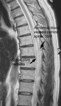
MRI scan showing features of venous hypertension |
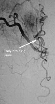
Selective angiogram of an intercostal artery |
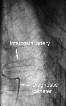 Unsubtracted image showing catheter in position |
|
In selected cases of spinal DAVFs and perimedullary fistulae, embolisation may provide a definitive cure without the need for open surgery.
Pre-operative embolisation may also be sought as a prelude to the resection of spinal tumours. Some cancers such as renal and thyroid carcinomas commonly spread to the spine and are typically extremely vascular. Pre-operative embolisation can significantly reduce the amount of intraoperative blood loss.